Highly Detailed Female Pelvic Model (Magnetic)


Highly Detailed Female Pelvic Model (Magnetic)
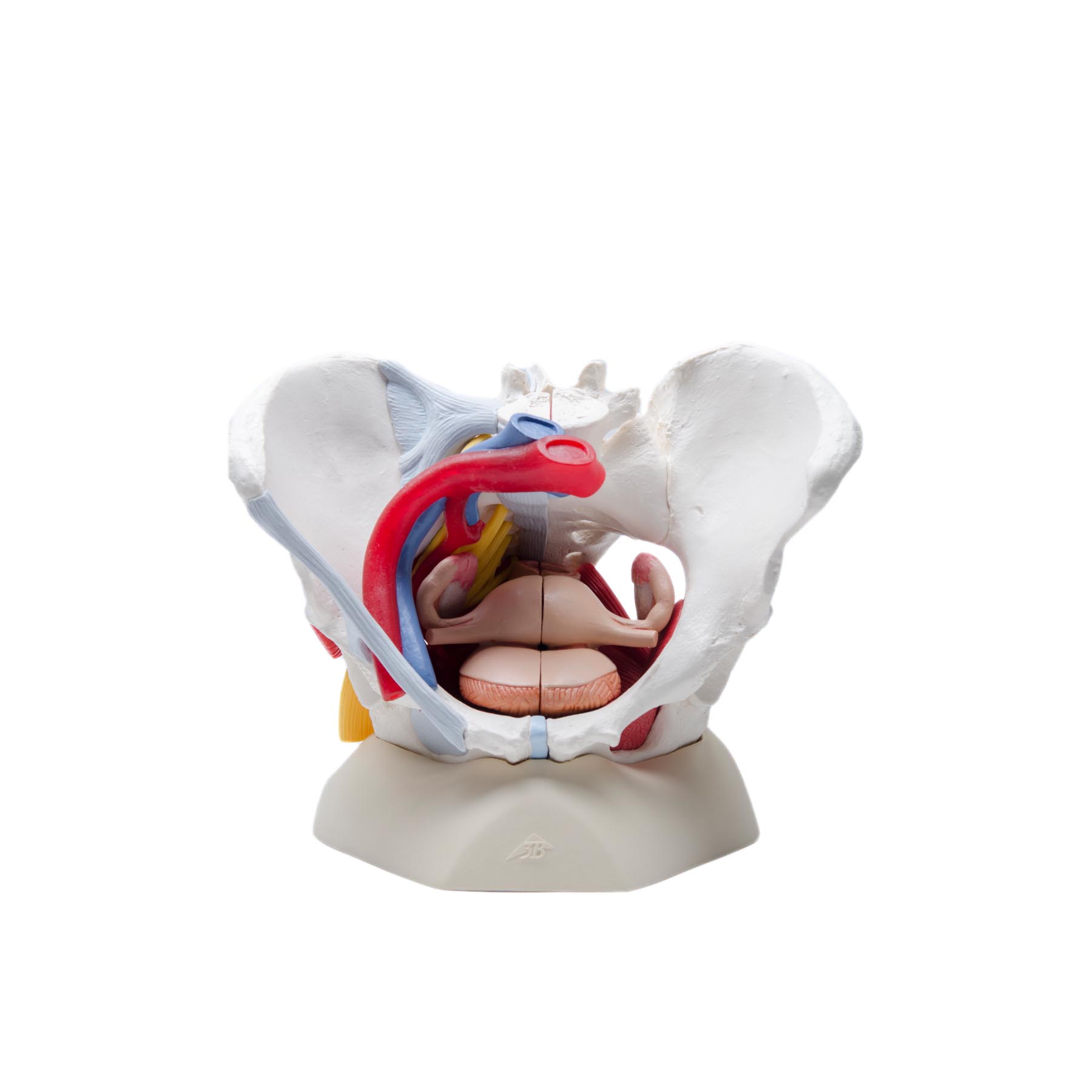
SKU: AS-H204This life size six-part female pelvic model provides detailed information about the topography of bones, ligaments, vessels, nerves, pelvic floor muscles and female genital organs.
It presents the whole pelvic floor with partially removable mid-sagittally sectioned external anal sphincter, external urethral sphincter, deep and superficial transverse perineal and bulbospongiosus.
What does this model offer?
This life size six-part female pelvic model provides detailed information about the topography of bones, ligaments, vessels, nerves, pelvic floor muscles and female genital organs. It presents the whole pelvic floor with partially removable mid-sagittally sectioned external anal sphincter, external urethral sphincter, deep and superficial transverse perineal and bulbospongiosus.
The rectum, uterus with fallopian tubes, ovaries and vagina of the female pelvic model are also removable and can be disassembled into both halves by midsagittal section. The right pelvic half demonstrates the divisions and topographical anatomy of the common iliac artery, the external and internal artery and also of the common iliac vein and the external iliac vein. The right sacral plexus, right sciatic nerve and right pudendal nerve are also shown.
Bones and ligaments presented: Two hip bones, the pubic symphysis, the sacrum and the coccyx, the fifth lumbar vertebra with intervertebral disc. A midsagittal section through the fifth lumbar vertebra, sacrum and coccyx, allow both halves of the pelvis to be disassembled revealing a part of the cauda equina in the vertebral canal.
The left half of the fifth lumbar vertebral body is removable. The right half of the model shows the following pelvic ligaments: inguinal ligament, sacrotuberous ligament, sacrospinous ligament, anterior sacroiliac ligaments, iliolumbar ligament, anterior longitudinal ligament, interosseous sacroiliac ligament, posterior sacroiliac ligament and obturator.
This model is great for detailed study of the female genital and pelvic anatomy. Whether you’re trying to explain the use of dilators or pelvic floor massage tools the female pelvic model is an excellent choice for clinicians needing to explain procedures to patients prior to administration!
Testimonial:
I just have to say that I really love the biofeedback and the pelvic floor model that I have from CMT. I use them both with almost every patient. People have told me that the 3-dimensional model has really helped them to understand the pelvic floor better, for both men and women. And of course, everyone raves about the biofeedback as it really helps them to build confidence that they are doing their pelvic floor exercises correctly.
Diane E. Hubbard, PT
Mercy Health
| Weight | 5.01 lbs |
|---|---|
| Dimensions | 12.13 × 10 × 12.13 in |

Female Genital Organs Anatomy Chart
The Female Genital Organs Anatomy Chart is an excellent example of a beautifully illustrated and painstakingly labeled anatomical poster....
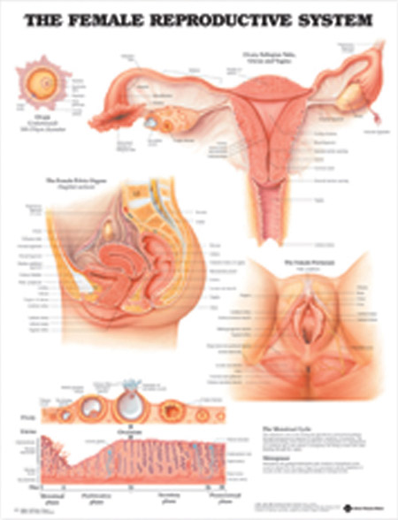
Female Reproductive System Anatomical Chart
The Female Reproductive System Anatomical Chart is an informative 20"x26" laminated chart that details the Female reproductive system to...